
CARING FOR LIVING ARTWORKS
Learn about the living cells and tissues in Emergence and the artist-biologists who maintained their health.

BJ-5ta immortalized human fibroblast, an A-375 Melanoma human fibroblast, an HC-11 mammary gland, and human lacrimal gland organoids in the Emergence laboratory CO2 incubator. Image credit: Fathomers
Living cells and tissues were central to several of the artworks in Emergence: Art from Life. Artist-biologists Maria Cabrera Abad and Kacy Jung were hired as Emergence lab technicians, and maintained the health of cell lines and organoids on a near-daily basis for the entirety of the exhibition's three-month run.

Examining tissue under the Emergence laboratory inverted microscope. Image credit: Fathomers
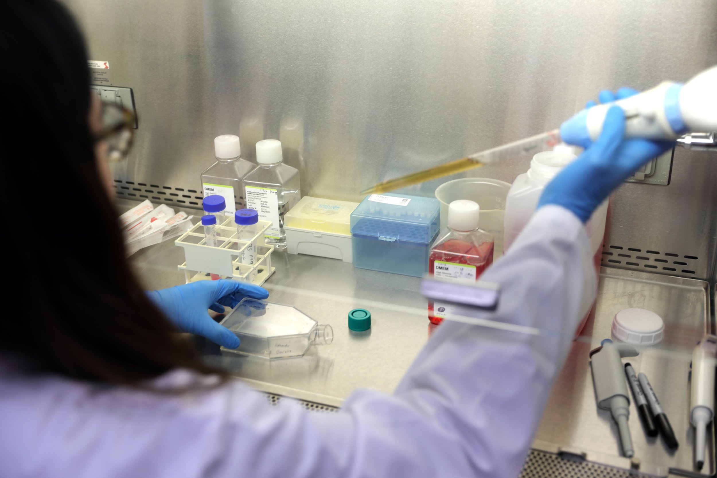
Maintaining cell health inside the Emergence laboratory biosafety cabinet. Image credit: Fathomers
Living materials were tended to in a specially created biosafety level 2 laboratory built out with a CO2 incubator, centrifuge, inverted microscope, biosafety cabinet, water bath, freezer, and other equipment. There, Maria and Kacy walked through a regimen of care for the most ancient, basic forms of life on Earth: the health of the cells would be examined underneath the microscope, the nutrient-rich media in which they rested would be changed, and the cells would be placed overnight in the incubator, which was kept at the mammalian body temperature of 37 degrees Celsius.

Emergence biosafety level 2 laboratory. Image credit: Carson Davis Brown

Emergence biosafety level 2 laboratory. Image credit: Carson Davis Brown
A careful record was maintained of the passage from one media to another by each cell line, which included a BJ-5ta immortalized human fibroblast, an A-375 Melanoma human fibroblast, an HC-11 mammary gland, and human lacrimal gland organoids.
The lab was utilized by various Emergence artists-scientists and visited by members of the public, who suited up in PPE, for a special behind-the-scenes tour of the lab and caretaking that made Emergence possible.
Emergence: Art from Life was made possible thanks to PST ART: Art & Science Collide presented by Getty.

Apoptotic Bodies (installation detail), Eduardo Padilha, 2024. Image credit: Fathomers

Apoptotic Bodies, Eduardo Padilha, 2024. Image credit: Carson Davis Brown

The Use of Life (in Relation to the Industry of Men), The Tissue Culture & Art Project: Oron Catts and Ionat Zurr, with assistance from UCLA Art|Sci and the California NanoSystems Institute, 2024. Image credit: Carson Davis Brown

Crying Organoids, Dr. Marie Bannier Hélaouët, Organoid Group, Hubrecht Institute of the Royal Academy of Arts and Sciences (KNAW), The Netherlands; Oncode Institute, The Netherlands; and Dr. Albert Wu, Ophthalmic Stem Cell and Regenerative Medicine Laboratory, Stanford Medicine, with lab members Dr. Aditi Swarup, Dr. Hala Dhowre, Dr. Sanja Bojic, and Julietta Picco, 2024. Image credit: Carson Davis Brown